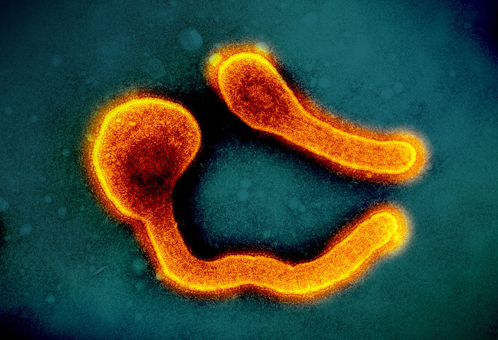
When Marburg virus (MARV) and Ebola virus (EBOV) erupt in human populations, their lethality is swift and devastating. Despite decades of research, treatments remain limited. Now, scientists from the Institute of Tropical Medicine at Nagasaki University (ITM-NU) in Japan, working with colleagues at the Philipps-Universität Marburg BSL-4 facility in Germany and other partners, have mapped in detail how these filoviruses assemble and transport their genetic material inside host cells. The study, recently published in mBio, offers critical molecular insights that could open doors to broad-spectrum antiviral strategies
Key Proteins Behind Viral Transport
Using advanced live-cell imaging, researchers identified three viral proteins—nucleoprotein (NP), VP35, and VP24—as both necessary and sufficient to form transport-competent nucleocapsid-like structures (NCLSs) in MARV. These structures shuttle viral genetic material through the cell to the plasma membrane, where new virions form. While additional proteins like polymerase L and VP30 are part of the viral complex, the study showed they are not required for nucleocapsid transport.
This finding mirrors prior observations in Ebola virus, underscoring a conserved mechanism across filoviruses. The actin cytoskeleton was shown to be critical: disrupting actin filaments halted nucleocapsid transport, while microtubule disruption had little effect.
Cross-Virus Compatibility and a Shared Weakness
The team also explored whether nucleocapsid proteins from MARV and EBOV could substitute for one another. They found that most were incompatible—except for VP30, a transcription factor. Strikingly, VP30 from either virus could partially function in the other, sustaining limited levels of replication in heterologous systems.
At the center of this compatibility is a small, conserved amino acid sequence in NP called the PPxPxY motif. This motif regulates NP’s interaction with VP30, enabling the protein to dock onto nucleocapsids. When the motif was mutated, VP30 could no longer bind, impairing transcription and preventing its incorporation into virus-like particles. Because this motif is conserved across the filovirus family, it emerges as an attractive candidate for broad-spectrum therapeutic targeting.
Looking Forward
MARV and EBOV continue to spark outbreaks in Africa. Antivirals remain scarce, and vaccines are limited primarily to Ebola. By identifying a shared vulnerability in the viral assembly line, the study highlights a potential path toward treatments that could apply across multiple filoviruses.
Further work with recombinant viruses, particularly VP30-deficient strains, will be necessary to confirm the motif’s role in natural infection. Moreover, understanding how host proteins interact with the PPxPxY motif could broaden its appeal as a drug target, but also complicate specificity.
Still, the study marks an important advance in decoding the inner workings of some of the world’s most dangerous pathogens. By revealing both commonalities and distinctions between Marburg and Ebola viruses, it provides a foundation for designing interventions that may finally blunt their deadly impact.
Takamatsu Y, Dolnik O, Hirabayashi A, et al. Molecular insights into nucleocapsid assembly and transport in Marburg and Ebola viruses. mBio. 22 September 2025


