

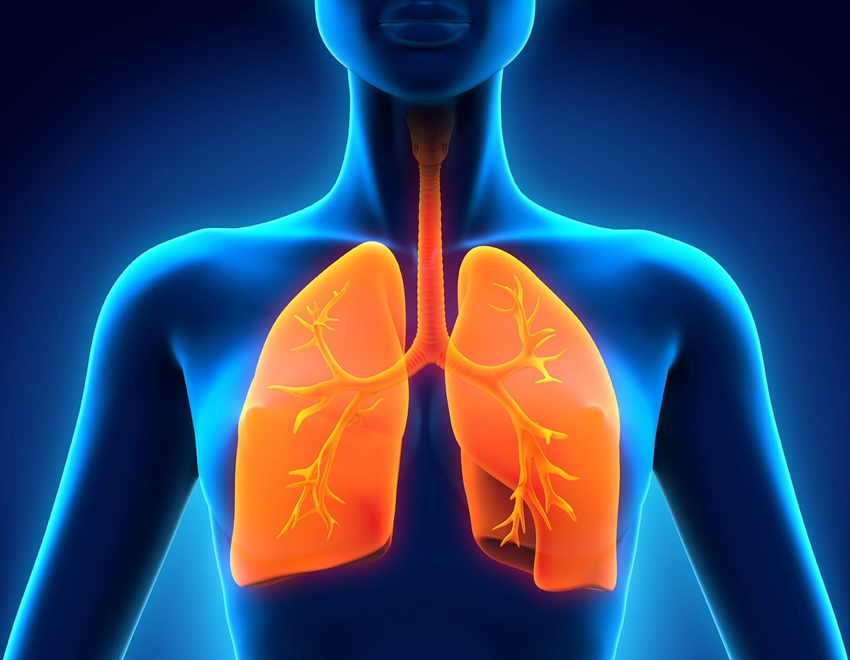
To the naked eye — or even on a chest X-ray — the scarred, obstructed lungs of a soldier exposed to sulfur mustard decades ago during the Iran-Iraq War can look strikingly similar to those of a lifelong cigarette smoker. Both show airway obstruction, tissue damage, and impaired breathing. But deep inside those damaged cells, a new study suggests, the biological machinery driving the injury is fundamentally different — a distinction that could reshape how doctors approach treating chemical weapons casualties.
Researchers from Iran’s Baqiyatallah University’s Chemical Injuries Research Center analyzed genetic activity in airway cells exposed to sulfur mustard, cigarette smoke, and other types of lung injury. Their findings, published in the Journal of Genetic Engineering and Biotechnology, suggest that the specific genes and cellular programs activated by mustard gas are distinct from those triggered by smoking — and even distinct from COPD, the chronic obstructive lung disease that smoking can cause. The work carries direct implications for developing treatments tailored to chemical weapons exposure, a concern for preparedness planners dealing with legacy populations of injured veterans and the persistent threat of chemical weapons use.
How cells repair themselves — and when that goes wrong
To understand the study, it helps to know how lung tissue normally responds to injury. When airway cells are damaged, they undergo a carefully choreographed biological dance: they can partially lose their identity as surface epithelial cells and temporarily adopt properties more like deeper tissue cells — a process called epithelial-to-mesenchymal transition, or EMT. This allows them to migrate and fill in gaps from the injury. Once healing is underway, they’re supposed to switch back to their original identity in a reverse process called mesenchymal-to-epithelial transition, or MET. In healthy lungs, this cycle of transition and reversal keeps remodeling controlled and tissue repair efficient.
In chronically damaged lungs — whether from mustard exposure or smoking — this cycle breaks down. The cells get stuck in an imbalanced state where they can’t fully complete either transition, leading to ongoing, ineffective tissue remodeling and progressive scarring. The researchers call this “repair imbalance,” and both mustard-injured lungs and smoking-related COPD show signs of it. But the study’s key finding is that they arrive at this state through different molecular routes.
What makes mustard different from smoke
The research team analyzed gene expression — the set of genes that cells activate or silence — across five groups: people with acute (recent) sulfur mustard exposure, those with chronic Mustard Lung disease from old exposures, cigarette smokers, COPD patients, and a lab-control group with mechanical airway injury as a reference.
When sulfur mustard hits airway cells, it causes chemical damage to DNA itself — bulky lesions and strand breaks in the genetic code. In response, the cells activate a suite of specialized DNA repair programs, including pathways specifically designed to fix those kinds of lesions. The study found that these DNA damage response pathways lit up robustly after acute mustard exposure but were largely absent in the other groups. This makes biological sense: mustard’s chemical mechanism directly injures DNA, triggering an emergency repair response that smoke inhalation doesn’t elicit in the same way.
Cigarette smoke, by contrast, was associated with different genetic activity: genes involved in maintaining telomeres — the protective caps at the ends of chromosomes that shorten with age and toxic stress — showed distinct patterns. Chronic smoke inhalation appears to accelerate the shortening of these telomeres, triggering a different set of cellular stress responses than what mustard does acutely. Interestingly, acute mustard exposure did show some telomere-related gene activity, but with a different signature than smoke, suggesting the cell is experiencing telomeric stress but processing it through a different biological pathway.
The researchers also identified specific “hub genes” — genes that act as command centers coordinating broader networks of genetic activity — that were activated in some conditions but not others. For instance, genes involved in fibrosis (tissue scarring), like fibronectin and a protein called SERPINE1, were upregulated in smoking and COPD but decreased after mustard exposure. Other genes, like ERBB2 and ERBB3, were preferentially activated after smoke exposure. The picture that emerges is one of condition-specific molecular signatures: the same end result — damaged, scarred lungs — produced by different cellular programs.
Why this matters for treating chemical weapons casualties
Sulfur mustard remains a documented chemical weapons threat with significant legacy populations — tens of thousands of Iranian veterans still live with Mustard Lung disease decades after exposure — and continued proliferation risk in certain regions. If the molecular pathways driving acute and chronic mustard injury are distinct, as this study suggests, then treatments might need to be phase-specific: drugs designed to boost DNA repair could be most effective when given soon after exposure, while interventions aimed at stopping tissue remodeling and fibrosis might be better suited to managing chronic sequelae years or decades later.
The identified hub genes represent potential drug targets. The key point for policy is this: chemical weapons injury has its own distinct molecular biology, which means that medical countermeasures — treatments, drugs, or interventions — designed for COPD or smoking-related disease may not work well for mustard-exposed populations. Dedicated research programs and drug development pipelines tailored to chemical casualty management, rather than borrowed from existing COPD frameworks, are likely needed.
All the analysis was computational (done by analyzing existing genetic datasets in a computer) rather than based on new experiments or patient studies. The datasets being analyzed were relatively small and used different laboratory methods, which can introduce noise. The researchers used statistical screening methods that could flag false positives without strict correction for multiple comparisons. Most fundamentally, the study looked at bulk gene expression in mixed cell populations, meaning it cannot pinpoint which specific types of cells within the airway are driving each response. The authors describe their findings as hypothesis-generating, requiring validation through targeted laboratory experiments, genetic studies in animal models, or culture systems that mimic airway tissue.
Sources and further reading:
Arabfard M, Salehi Z, Ghanei M, Azimzadeh Jamalkandi S. Transcriptomic landscape of airway epithelial repair: Contrasting acute and chronic injury in mustard lung and COPD. Journal of Genetic Engineering and Biotechnology. 24 June 2026.