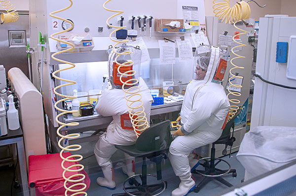
A new study published in npj Imaging sheds new light on the role of oxidative stress in Ebola virus disease (EVD). Conducted at the National Institute of Allergy and Infectious Diseases (NIAID) Integrated Research Facility (IRF) at Fort Detrick—one of the few federal labs authorized to study Ebola under biosafety level 4 (BSL-4) conditions—the research provides critical new data on how reactive oxygen species (ROS) contribute to immune cell loss and disease progression.
This vital research comes at a time of heightened concern, as the IRF has recently been shut down by the Trump administration, raising alarms in the global health community about the U.S. government’s ability to conduct essential high-containment pathogen research.
The study, led by Venkatesh Mani and colleagues at the IRF, uses a combination of ex vivo immunohistochemistry and in vivo ROS-sensitive MRI in domestic ferrets to establish a link between oxidative tissue damage and the depletion of T and B lymphocytes in the spleen—a hallmark of severe EVD.
ROS-Driven Tissue Damage Tracks with Disease Progression
Researchers observed a progressive increase in ROS-related oxidative stress markers in the spleens, livers, and kidneys of Ebola-exposed ferrets. Notably, the accumulation of 4-hydroxy-2-nonenal (4-HNE) and myeloperoxidase (MPO)—biomarkers for oxidative stress—was highest in the spleen at terminal stages of the disease. MRI imaging using the Fe-PyC3A probe confirmed these findings in vivo, detecting significant ROS-related signal enhancement corresponding to disease severity.
Splenic Lymphocyte Depletion Correlates with Oxidative Stress
Flow cytometry revealed a marked decline in CD4+ and CD8+ T cells in the spleens of ferrets as the disease progressed. CD4+ T cell apoptosis increased steadily, and this pattern significantly correlated with ROS-related tissue changes detected by both MRI and immunohistochemistry. B cells also showed increased apoptosis, although their absolute numbers remained stable. These findings suggest ROS may play a direct or indirect role in immune cell death during Ebola infection.
First Application of Molecular MRI to EVD in a Small-Animal Model
This study represents the first use of molecular MRI with a ROS-sensitive probe (Fe-PyC3A) in a small-animal model of Ebola. The approach allowed for longitudinal, non-invasive imaging of oxidative tissue changes, opening up new possibilities for studying immune dysregulation and therapeutic intervention in high-containment environments.
Evidence of Systemic Immune Disruption
Beyond splenic findings, the study confirmed systemic immune cell loss and elevated serum cytokines, including TNF—a cytokine known to amplify ROS production. These results align with known immune collapse patterns in human and nonhuman primate cases of EVD, reinforcing the model’s translational relevance.
Implications for Future Research and Preparedness
The data strongly support a role for ROS in the pathogenesis of EVD and raise new questions about whether therapies targeting oxidative stress could mitigate immune cell loss and improve outcomes. Importantly, the study’s success underscores the critical capabilities of BSL-4 facilities like the now-closed IRF in Frederick, Maryland.
Given the challenges of safely studying lethal pathogens, the use of ferrets and molecular imaging in this study provides a scalable model for future investigations. It also highlights the irreplaceable role of federal research infrastructure in preparing for and responding to biological threats.
About the BSL-4 Facility
The study was conducted at the Integrated Research Facility (IRF) at Fort Detrick, part of the National Institute of Allergy and Infectious Diseases (NIAID). The IRF was among the few BSL-4 labs in the United States capable of safely conducting advanced Ebola virus research. Its recent closure, as reported by Wired, has sparked concern about the nation’s long-term preparedness for handling the world’s most dangerous pathogens.
Mani, V., Chu, W.T., Yang, H.J., et al. (2025). Reactive oxygen species-related oxidative changes are associated with splenic lymphocyte depletion in Ebola virus infection. npj Imaging, 3, 16. https://doi.org/10.1038/s44303-025-00079-x


