

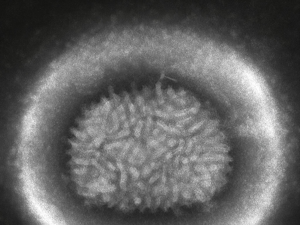
Neurological complications following vaccinia virus infection have been documented since the era of mass smallpox vaccination. Encephalitis, postvaccinal encephalopathy, and less severe sensory disturbances were among the rare but serious adverse events associated with live replication-competent vaccines. Yet the mechanisms by which vaccinia virus reaches and damages the central nervous system have remained poorly characterized, in part because suitable animal models have been lacking. A new study published in Frontiers in Immunology fills that gap, using an intranasal mouse infection model to trace orthopoxvirus neuroinvasion from the nasal epithelium to the brain and documenting the consequences for olfactory function in real time.
The study, conducted by researchers at the Shanghai Public Health Clinical Center, Fudan University, and Shanghai Normal University, used VACV VR-1354, a tissue culture-adapted derivative of the neurovirulent Western Reserve vaccinia strain, to infect two mouse strains via the intranasal route. The work is the first to comprehensively characterize the virological, histopathological, transcriptomic, and behavioral consequences of olfactory-route orthopoxvirus neuroinvasion using this model.
A Direct Highway to the Brain
The study’s foundational finding is that VACV VR-1354 efficiently disseminates from the nasal mucosa to the brain following intranasal inoculation, following a spatiotemporal gradient that implicates the olfactory epithelium as the primary entry point. Viral DNA was detectable in the nasal mucosa as early as three days post-infection, peaking at seven days and remaining elevated at day 14. Viral loads in the olfactory bulb followed a similar but lagged pattern, while the cerebrum and cerebellum showed only low-level viral DNA at the seven-day peak, with near-absent signal at days three and fourteen. The distribution pattern, with nasal mucosa highest, olfactory bulb intermediate, and cerebrum and cerebellum substantially lower, is consistent with direct neuroinvasion via the olfactory nerve rather than hematogenous spread from the systemic circulation.
C57BL/6N mice exhibited significantly higher viral loads at peak infection than BALB/c mice, correlating with greater weight loss and higher mortality (41.7% versus 30.0%) and with more severe downstream neuropathological findings.
Blood-Brain Barrier Disruption
To assess the integrity of the blood-brain barrier, the researchers used Evans blue dye extravasation, a standard method for detecting vascular permeability, at three, seven, and 14 days post-infection. Results showed a clear temporal pattern: minimal dye penetration at day three, maximal leakage in the olfactory bulb at day seven, and resolution by day 14. The effect was more pronounced in C57BL/6N mice than in BALB/c mice, consistent with the strain differences in viral load and disease severity.
The finding that BBB disruption is both localized to the olfactory bulb and transient is significant. It suggests that orthopoxvirus-induced CNS pathology need not involve systemic vascular compromise and that recovery of barrier function is possible. As the behavioral data show, however, functional consequences can persist well beyond the resolution of structural injury.
Reversible Chemosensory Loss
The most clinically translatable finding concerns olfactory behavior. The researchers assessed chemosensory function in C57BL/6N mice using a camphor aversion assay. Uninfected mice reliably prefer to spend time in an odor-free chamber over one containing camphor solution, a preference that depends on intact olfactory function. Infected mice showed progressive erosion of this aversion preference beginning at 14 days post-infection and persisting through 49 days post-infection. From days 28 to 49, infected mice showed no significant preference for the odor-free side, indicating substantial chemosensory impairment. Functional recovery was observed by day 56.
The authors note that this recovery timeline, extending more than five weeks beyond the resolution of both viral load and acute neuroinflammation, implies that chemosensory function requires prolonged olfactory epithelium reinnervation and synaptic remodeling in the olfactory bulb, rather than simply clearing the virus and resolving inflammation.
Post-Viral Olfactory Loss and Orthopoxvirus Preparedness
The parallels to post-COVID olfactory dysfunction are explicit in the paper’s framing. The combination of olfactory sensory neuron injury, olfactory receptor gene downregulation, transient BBB disruption, and delayed functional recovery recapitulates key features of post-viral anosmia regardless of the specific pathogen involved. The VACV VR-1354 mouse model demonstrated here offers a genetically tractable and mechanistically accessible platform for studying these processes.
With mpox documented to invade the CNS via the olfactory route in recent studies, and with a global population increasingly naive to orthopoxviruses following the end of smallpox vaccination, understanding the neuropathogenic mechanisms of this virus family has renewed urgency. This model provides a platform for evaluating potential therapeutic interventions targeting both viral neuroinvasion and sensory recovery — an area where no approved treatments currently exist.
Sources and further reading:
Li S, Wu Y, Wang C, et al. Intranasal VACV VR-1354 infection impairs chemosensory function and induces olfactory bulb neuroinflammation in mice. Frontiers in Immunology, 19 April 2026