

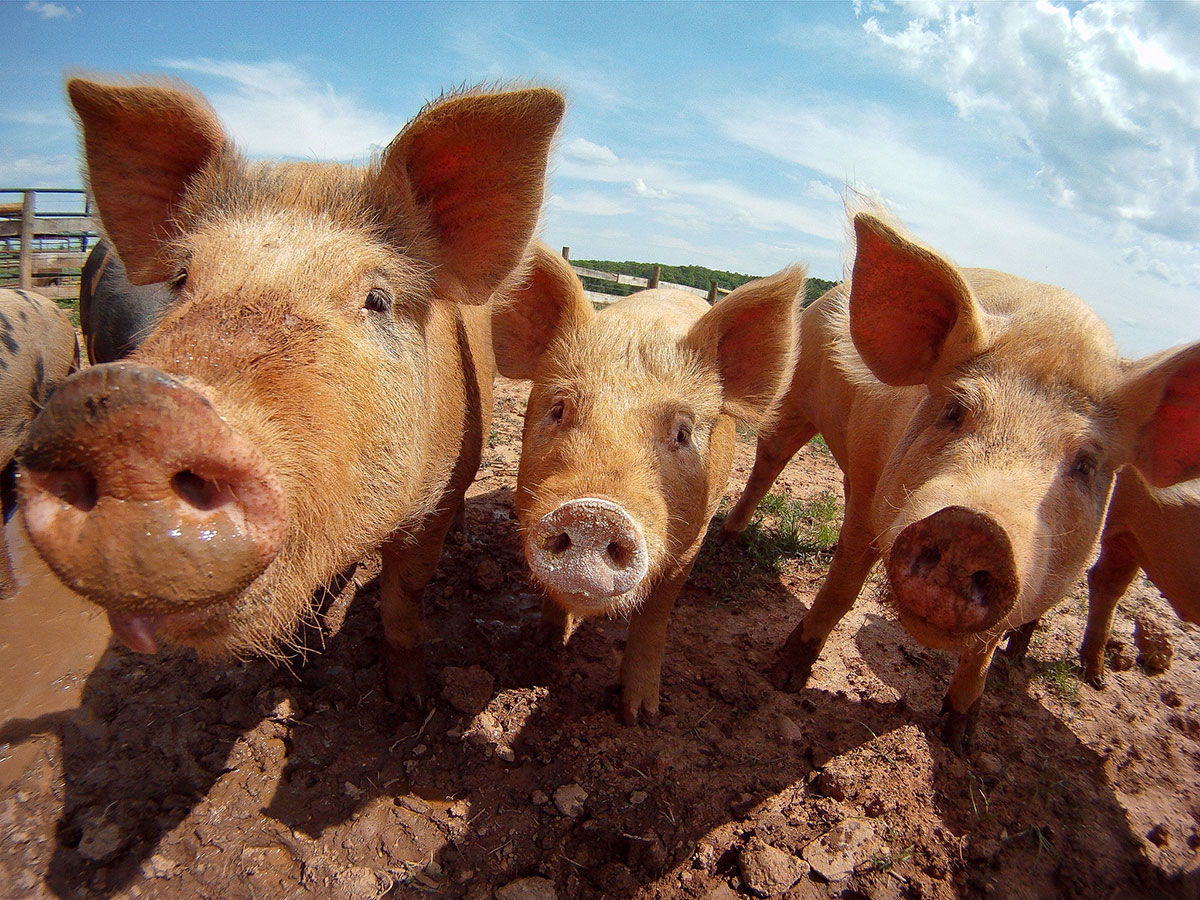
Researchers experimentally inoculated pigs with H5N1 genotype D1.2, a strain first detected in a backyard sow in Oregon in October 2024, and documented how the virus behaves in mammalian hosts. The study reveals a troubling pattern: the infected animals showed no fever, no respiratory symptoms, and no overt illness, yet the virus replicated efficiently in nasal secretions for multiple days and spread systemically to the brain, muscle tissue, lymph nodes, and digestive organs. Notably, researchers recovered active virus from the diaphragm muscle itself, suggesting that meat from infected animals could potentially pose a contamination risk.
The genotype D1.2 emerged through reassortment between the dominant D1.1 strain circulating in North American birds and low-pathogenicity avian influenza viruses. Genetic analysis shows D1.2 carries multiple mammalian adaptation markers, including mutations in viral polymerase genes associated with increased replication in mammalian cells and enhanced virulence in animal models. These genetic characteristics may explain the virus’s ability to establish efficient infection in swine despite the species barrier that typically limits avian influenza replication in mammals.
Throughout the 35-day study period, infected pigs shed virus consistently in nasal secretions, suggesting they could transmit H5N1 to other animals or potentially to handlers in close contact. Yet because the animals appeared healthy and exhibited no respiratory distress, illness in commercial settings would be easily missed. This detection challenge is compounded by a weak immune response. The infected animals developed minimal to no neutralizing antibodies against the virus, meaning standard serological testing for H5N1 exposure would not reliably identify infected animals after recovery.
The systemic spread was extensive. At five days after infection, researchers detected viral antigen and live virus in tissues spanning the respiratory tract, brain, skeletal muscle, lymph nodes, and digestive system. The virus infected multiple cell types, including neurons in the olfactory bulb and cerebral cortex, alveolar macrophages in the lungs, and various immune cells in lymph nodes. The detection of viral antigen in skeletal muscle is particularly significant given that diaphragm tissue had infectious virus present at levels detectable by standard laboratory methods.
By day 35 post-infection, virus persisted in the brain and upper respiratory tract of some animals despite minimal clinical illness. This protracted, subclinical infection suggests that even recovered animals could harbor infectious virus in tissues for extended periods. The ability to detect viral antigen deep within the brain tissue weeks after inoculation indicates that H5N1 D1.2 can establish persistent infection in the central nervous system, a finding consistent with the known neuroinvasive capacity of highly pathogenic H5N1 strains.
The findings point to a longstanding concern in influenza surveillance: swine serve as potential mixing vessels for reassortment between avian, human, and swine influenza viruses. These animals express sialic acid receptors on their respiratory epithelium that permit binding of both avian-type and mammalian-type influenza viruses. If H5N1 establishes circulation in commercial swine while producing minimal clinical signs, the virus could spread undetected and potentially reassort with endemic swine influenza viruses, generating novel strains with altered characteristics.
For food safety, the isolation of infectious virus from meat juice raises questions about the adequacy of current slaughter and processing procedures to eliminate H5N1 contamination. Standard cooking temperatures do inactivate influenza viruses, but the presence of viable virus in meat at the point of processing indicates that contamination is possible. If infected animals enter slaughter facilities undetected due to lack of clinical signs, virus-contaminated carcasses could potentially reach consumers.
The weak antibody response is particularly troubling for herd surveillance. Serology is often used to identify exposed or infected animals after an outbreak, but these findings suggest that H5N1-infected swine may not develop robust detectable antibodies. This means that animals identified as negative by serological testing could still be infected or recently recovered from active infection.
The study was conducted under controlled conditions with a specific virus dose and route of inoculation. Commercial infections may follow different trajectories depending on exposure level, environmental stress, co-infections, and other variables. However, the findings from this experimental work represent a realistic scenario given that D1.2 is already circulating in backyard and potentially commercial swine holdings in the United States. As the H5N1 outbreak in dairy cattle continues and the virus spreads geographically, spillover into commercial swine herds becomes an increasing risk.
Early detection of any H5N1 circulation in herds will require regular testing of apparently healthy animals, not reliance on clinical signs or post-infection serology alone.
Sources and further reading:
Seger H, Baker AL, Buckley AC, et al. Detection of Highly Pathogenic Avian Influenza A(H5N1) Clade 2.3.4.4b Genotype D1.2 Virus in Swine after Experimental Inoculation. Emerging Infectious Diseases, July 15, 2026.
This article was researched and sourced by Global Biodefense editors and reported with Claude AI assistance for drafting and editing.